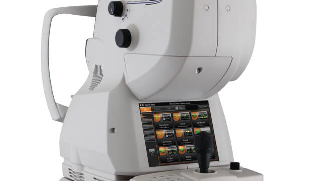
The DRI OCT Triton combines the world’s first Swept Source OCT technology with multimodal fundus imaging.
Key features
1,050nm wavelength
100,000 A-scan/sec scanning speed
Multimodal fundus imaging
Image Quality
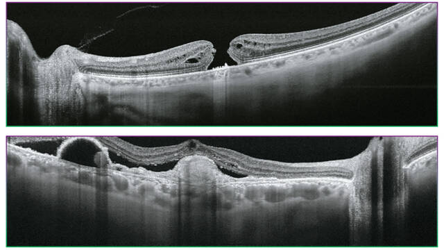
Triton’s Swept Source with its 100 kHz scanning speed and 1,050nm wavelength results in a clear and detailed images even for the deepest layers of the eye with short acquisition time. Visualize not only the retina and vitreous, but also the choroid and sclera1.
Diagnostic capability
Seeing deeper makes it possible to have a better understanding of many ocular pathologies1.With features such as OCT angiography, fundus autofluorescence and en face OCT, Triton empowers clinicians with multimodal imaging capability to help assess and preserve patient’s eye health.
Practice efficiency
The Triton’s automated functions, including single scan captures and SMARTTrackTM system, are designed to optimize your practice workflow by simplifying data capture, analysis and follow-up.
1 Fabio Lavinsky, Daniel Lavinsky. Novel perspectives on swept‑source optical coherence tomography. Int J Retin Vitr (2016) 2:25
Courtesy: Professor Jose Maria Ruiz Moreno MD, University of Albacete, Spain
Swept Source OCT Imaging
1,050nm wavelength
The longer wavelength light provides better tissue penetration, allowing visualization into the deepest layers of the eye1
Swept Source OCT technology; scanning speed of 100,000 A-scans/sec
The fast scanning speed of 100,000 A-scans/sec enables capture of clear B-scans* by acquiring more A-scans within a given image acquisition time. This helps to reduce artifacts from involuntary eye movements such as saccades and blinks. *Shoji Kishi. Impact of swept source optical coherence tomography on ophthalmology. Taiwan Journal of Ophthalmology 6 (2016) 58-68
Invisible scan lines
The invisible 1,050nm wavelength light helps patients concentrate on the fixation target during the scan, reducing involuntary eye movement. It supports more efficient workflow in a practice by reducing the need to rescan.
Swept Source OCT incorporates multimodal fundus imaging
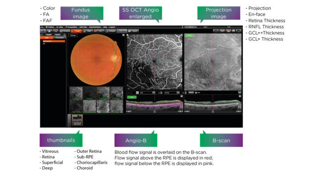
DRI OCT Triton can acquire the OCT and fundus image in a single capture. PinPointTM Registration identifies the location of the B-scan on the fundus image. Comparison between the B-scan and fundus image can support clinical efficiency during diagnosis.
True color* Fundus images
The DRI OCT Triton offers a true color, non-mydriatic fundus image. Fluorescein Angiography (FA) and Fundus Autofluorescence (FAF) are available** to enhance the diagnostic capability of Triton Plus. The all-in-one device supports efficient workflow in practice. Swept Source OCT incorporates multimodal fundus imaging DRI OCT Triton can acquire the OCT and fundus image in a single capture. PinPointTM Registration identifies the
location of the B-scan on the fundus image. Comparison between the B-scan and fundus image can support clinical efficiency during diagnosis. *Color fundus image with white light, with 24-bit color.
**DRI OCT Triton Plus :OCT /Anterior OCT (Option)/ OCT Angiography (Option) /Color /Red-Free / FA / FAF
DRI OCT Triton :OCT /Anterior OCT (Option)/ OCT Angiography (Option) /Color/Red-Free
Stereo photography
Three dimensional visualization of color fundus images can be achieved by acquiring images in stereo photography mode. Triton’s on-screen acquisition guidance supports quick and easy operation with auto alignment for capturing stereo pairs.
Panoramic wide field photography
In addition to macular and disc imaging, the Triton provides wide coverage of the retina. A panoramic graphic can be created from multiple fundus or OCT Angiography images.
Follow-up Function
This function allows you to retrieve and re-analyze the same location at follow-up, for comparison of past and current images. All an operator needs to do is simply select the past data and Triton automatically captures the same area.
Multimodal Viewing
En face angiography images, B-scans and fundus photography can all be viewed on a single screen using IMAGEnet®6 and PinPointTM registration, so that area of interest can be assessed using multiple image modalities. Selected layers can easily be customized to enhance the clarity of specific pathological features.
Discover more possibilities: see beyond and deeper
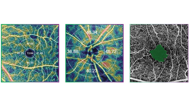
Topcons SS OCT Angio
Topcon’s SS OCT AngioTM combines OCT angiography with a Swept Source OCT. OCTARATM, a proprietary image processing algorithm, provides highly sensitive angiographic detection2 allowing for visualization of vascular structures even in the choroid and deeper retinal layers. 2Magdy Moussa, Mahmoud Leila, Hagar Khalid. Imaging choroidal neovascular membrane using en face swept-source optical coherence tomography angiography.
Clinical Ophthalmology 2017:11 1859–1869
OCTARATM
OCTARATM is the image processing technology which extracts the signal changes derived from vascular flow using multiple OCT B-Scans acquired at the same position. It demonstrates high sensitivity for the detection
of low blood flow in microvasculature2.
High-sensitivity Imaging and Deeper Intravascular Flow Visualization1
Swept Source technology and OCTARATM allow the deeper structures to be visualized with less depthdependent signal roll-off3. Additionally, the 1μm wavelength makes OCT Angiography imaging possible for patients with media opacities.
Rapid Scanning, Real Time Eye Tracking
At 100,000 A-Scans per second coupled with invisible* scanning lines and the SMARTTrackTM eye tracking system, ‘the Triton quickly captures a dense data set and provides an en face OCT Angiography image of
the retinal microvascular flow network’2 * OCT Angiography scanning line may be faintly visible during capture to some people with certain conditions
OCTA is optional extra.
SS OCT AngioTM Montage, Courtesy:Yuichiro Ogura, MD, Professor and Chairman, Department of Ophthalmology and Visual Science, Nagoya City University, Nagoya, Japan
Not all products, services or offers are approved or offered in every market, and products vary from one country to another. Contact your local distributor for country-specific information.
Triton Clinical Cases
Triton clinical cases
Triton OCT interpretation tips –
Luis Arias, MD, Ph.D., Head of Retina Department of University Hospital of Bellvitge and Aggregate Professor of O...
Clinical advantages: The reasons why DRI OCT Triton™ and OCTA Triton from Topcon Healthcare are the technologies of choice for retina and uveitis specialists
Outstanding Image Quality in a Fraction of the Time: Leading retina specialists share their impressions of PixelSmart™, the latest image processing algorithm from Topcon Healthcare